agalactiae antibodies in serum from 9th day of infection in accordance with results obtained by Fuscoet al. using immunoblotting. Colloidal gold nanoparticles were prepared from an aqueous chloroauric acidsolution (0.01%) by citrate reduction method . Salt agglomeration test was performed for determining the minimal protective amount and 10 μg of protein antigen mL was found to be protecting GNPs from salt agglomeration (Fig. 2). Further, the conjugation and blocking steps were also confirmed spectrophotometrically as increase in the absorbance of gold nanoparticles was observed after each step.
The stability of colloidal solutions for C-GNPs and their conjugated derivatives depended significantly on their size. Visible precipitates occurred for the average diameter of C-GNPs, which was equal to 47.5 after one to two months of storage . This effect may create worse sensitivity in the assay with these GNPs as a label. This finding is in accordance with earlier presented data about C-GNPs for large diameters that needed additional surface modifications to provide stability . The S-GNPs conjugated with antibodies possess long-time stability of colloidal solutions based on spectral and DLS data in a range of diameters up to 64.5 nm.
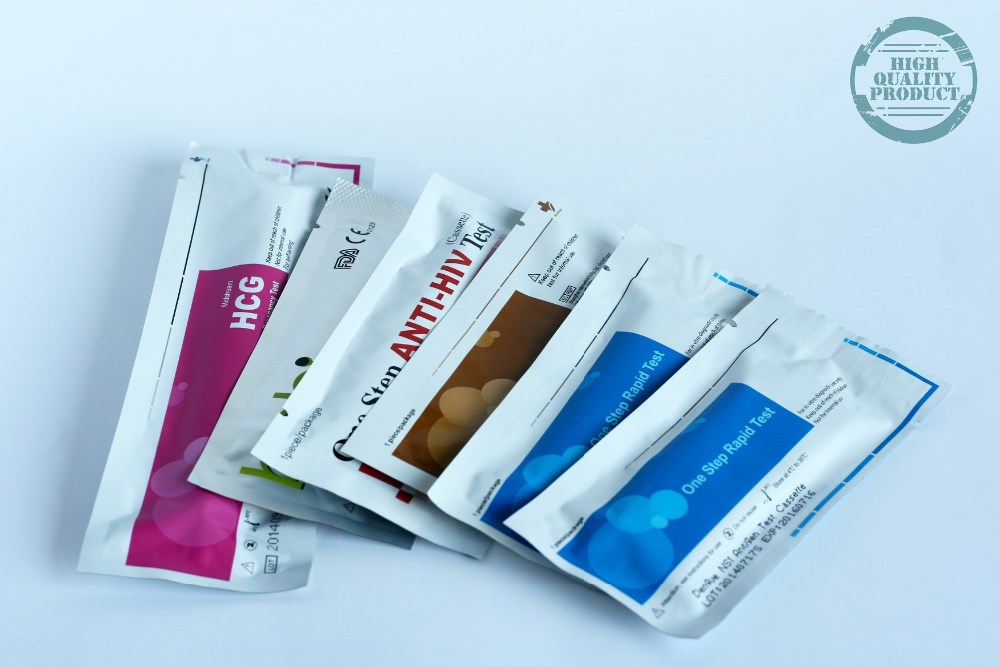
Quantitative Tests
This dedication to quality translates to dependable and consistently superior assay results. It also ensures better control of gold nanoparticle surface area and consistent flow dynamics across membranes along with high sensitivity from even binding of antibody or multiplexing modifications. The quality of gold nanoparticles can have profound effects on the specificity, sensitivity and reproducibility of lateral flow assays. Ideal for development of protein gold conjugates for use in applications such as blotting, lateral flow assays, microscopy and transmission electron microscopy . We are the only company in the world to offer Spherical Gold Nanoparticles from 1nm to 1500nm in diameter, Gold Nanorods with Surface Plasmon Resonances from 550nm to 2100nm, and Gold Nanowires up to 40 microns in length.
Apparently, a final comparison of the two options for immobilization is possible only for a significantly wider range of drugs, including antibodies to different antigens. LoDs of cTnI detection for LFIAs with different antibody–GNP conjugates. Based on the obtained concentration dependencies, the LoD values were determined for all four series of the conjugated GNPs, namely adsorption and covalent conjugates of C–GNPs and S-GNPs. To estimate the efficiency of visual assessment of the assay results based on the intensity of TZ coloration, the maximal saturating levels of these colorations for all tested kinds of GNP–antibody conjugates are summarized in Table 7. Dependencies of the intensities of staining of the test zone on the concentration of antigens in the sample for conjugates C-GNPs-1–C-GNPs-5 and S-GNPs-1–S-GNPs-5 during adsorption and covalent immobilization of antibodies. C-GNP and S-GNP series were synthesized with varied ratios of reactants to reach different average nanoparticle diameters. All of them were stable colloidal suspensions of red color, which is typical for nanodispersed gold.
Government Links
Although similar assays can be also designed using antibodies, aptamer sensors offer stability and low-cost advantages. Besides, aptamers are more flexible for developing different formats since they are composed of nucleic acids having intra- and inter-molecular hybridization, enzymatic replication, and easy sequence determination characteristics. In virtue of these positive properties, numerous aptamer sensors have been developed for multiplexed assays. Lateral flow strip assay was first developed in 1956 as a logical extension of the latex agglutination test technology . In view of the high occurrence of food security affairs and the common use of rongalite as an illegal food additive, it is necessary to develop an aptamer-based LFSA for the on-site and rapid detection of this compound in food samples. Once soaked, the fluid flows to the second conjugate pad in which the manufacturer has stored freeze dried bio-active particles called conjugates in a salt-sugar matrix. The conjugate pad contains all the reagents required for an optimized chemical reaction between the target molecule (e.g., an antigen) and its chemical partner (e.g., antibody) that has been immobilized on the particle's surface.
- The assay can be initiated by a simple contact of the test strip with the sample and does not require additional manipulations with reagents and devices.
- jirovecii levels results across patients with PcP and patients without P. jirovecii infection.
- The mechanism of passive adsorption is based on van der Waalsand other attractive forces between the antibody and the surface of the particle.
- The absence of the test line in the presence of the control line indicates a negative sample.
- Meanwhile, the maximum absorption peak exhibited a significant red shift from 527 nm to 598 nm with the color of AuNP solution changing from wine red to brick red with increasing AuNP size .
However, when the size of AuNPs exceeds 80 nm, the Qext of AuNPs mainly contributes to the increase of Qsca, whereas Qabs changes slightly . Previous work implied that the light absorption rather than scattering of AuNPs dominated the signal readout on the NC membrane .
Ultimately, this study will help in the management of PcP in industrialized countries, also having a major impact on developing countries with low income and lack of technology, where PcP is an emerging disease with high prevalence and poorly controlled. By visual inspection, it was observed that all NM can be used successfully without a previous blocking step. Additionally, the signal in the control and test lines appeared to increased proportionally with pore diameter and the wicking time of the NM. However, as membranes type CNPH are presented by the manufacturer as the NM with the highest protein binding capacity, the one with the longest wicking time was the one chosen for the LFIA development. The blocked and unblocked AuNP-RSA conjugates were further assessed by AGE to characterize their ability to interact with human sera from patients with and without P. jirovecii infection.
If no analyte exists in the test solution, then the reporter binds to the strip indicating a negative test. Fundamental to the performance of a lateral flow assay are the affinity reagents that recognize the biological target, utilized on both the particle and the test strip itself. Antibodies are a common choice that are sensitive and selective for the specific detection of very low concentrations of analyte. The AgraStrip® Total Milk kit is a ready-to-use lateral flow device supplied with all the consumables required to run 10 tests. Milk residue can be detected at any stage of the production process from testing surfaces, ingredients right through to finished product analysis.
Gold Nanoparticle
These signals have low background noise since there are generally no magnetic materials in the environment or in the tested samples. The study confirmed the Anteo Mix&Go based hCG assay is significantly more sensitive than the covalently conjugated magnetic particle based test using the same critical reagents. The hCG assay conducted with the Anteo Mix&Go particles used half the amount of antibody to achieve five times more sensitivity than the covalently conjugated assay. The limit of detection for the Anteo Mix&Go assay was ~25 mIU/mL in urine for the visual and reader based results. The limit of detection for the covalently conjugated hCG assay was ~100 mIU/mL in urine. As a reference, the limit of the detection for the colloidal gold assay is at ~25 mIU/mL in urine.
All rights reserved The Wick • Its task is to soak the sample liquid and all reagents that have not been absorbed at the test and control lines. • It must prevent the backflow of this liquid into the drying membrane as long as possible. • Select a cotton linters paper with an absorption capacity that is much higher than the sample volume. All rights reserved The Analytical Membrane • Typically, this is a “large pore sized“ nitrocellulose membrane. • The membranes are available in a very broad range of sample flow characteristics. • All NC membranes contain a surfactant, usually an anionic surfactant, that makes them hydrophilic. All rights reserved Conjugate Pad Materials • Options are glass fiber pads and non-wovens.
The mechanism of adsorption is based on van der Waals interactions between the proteins (e.g. antibodies) and the surface of the glass strip cutter particles. The resulting forces between the antibody and the nanoparticle are influenced by the coupling environment.
Alexandria Engineering Journal Impact
The test can be performed using 1 ml of venous blood, erythrocyte lysis buffer, a tabletop centrifuge, and a 37°C incubator without CO2. Although such a test cannot be used at the bedside, results with excellent precision and reliability and minimal laboratory capacity are available at 48 h. Our previous data suggest that a reading at 24 h may also be informative . We calculated the sensitivity, specificity, positive predictive value , and negative predictive value of the IgG LPS-specific lateral-flow dipstick using OpenEpi, version 3, an open source calculator for the evaluation of diagnostic tests.
agalactiae and protein concentration of antigen was adjusted to 2 mg mL-1using 0.01 M PBS. Contagious agalactia is an economically important disease of small ruminants which cause mastitis, agalactia, arthritis, keratoconjunctivitis, pneumonia and neonatal mortality (Bergonier et al., 1997).
Sonicated antigen prepared from standard and Indian strains was used as the capture probe in the assay. Recently, an indirect ELISA assay utilizing sonicated antigen (ELISA-Gs) of M. agalactiae was standardized for the serodiagnosis of contagious agalactia in goats (Campos et al., 2009). The sonicated antigen based ELISA showed sensitivity and specificity of 88.63 and 95.24%, respectively. The current lateral flow assay using sonicated antigen do not show any cross reactions with Mycoplasma mycoides ssp.,capri a Mycoplasma spp., that produce symptoms similar to that produced by M.
Winning The Contamination Control Battle
Under the optimum conditions, a series of HBsAg standard solutions in artificial serum with target concentration ranging from 0 ng/mL to 1000 ng/mL were tested simultaneously using AuNP40-and GSP270-LFIA strip. The strip photographs obtained at different HBsAg concentrations are shown in Figure 5A. The results indicated that the vLOD of GSP270-LFIA strip for HBsAg reached up to 0.46 ng/mL, which was ca. Figure 5B presents that the ODT/ODC value increased as the HBsAg concentration increased, and an excellent linear relationship between them was observed from 0.46 ng/mL to 1000 ng/mL with an R2 of 0.9902. The specificity analysis in Figure 5C suggested the excellent selectivity of this GSP270-LFIA strip for HBsAg against other common serum interferences, including AFP, CEA, HCG, PCT, HCV-Ab, and HSA.